04/20/1896 • 6 views
A First Glimpse: Early Public X-ray Demonstration in 1896
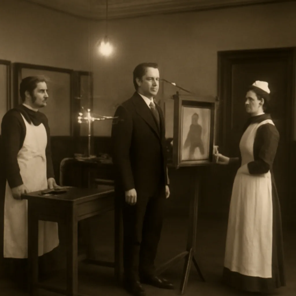
In April 1896, weeks after Wilhelm Röntgen’s discovery, public demonstrations of X-ray imaging began to appear in Europe and North America, revealing bones and foreign objects and immediately captivating scientists, physicians and the general public.
These demonstrations varied in setting and scale. In some cases they occurred in medical schools or hospitals, where physicians tested X-rays’ potential for diagnosing fractures and locating bullets; in others they took place in lecture halls, photographic studios or fairgrounds, where operators presented skeletal images to paying audiences. Early apparatus typically consisted of Crookes or Geissler tubes powered by induction coils, hand-wound spark coils, or static machines; exposures could be long by modern standards, ranging from seconds to many minutes, and photographic plates or fluoroscopic screens were used to capture or view the images.
Contemporary accounts emphasize both scientific curiosity and spectacle. Medical practitioners quickly recognized clinical uses, reporting cases in which X-rays helped identify fractures, locate swallowed or lodged metallic objects, or reveal foreign bodies. At the same time, newspaper reports and public lectures often highlighted the novelty and wonder of seeing “inside” the living body. Because photographic reproduction and press coverage spread rapidly, demonstrations in one city inspired others nearby; this diffusion explains why many places claimed early demonstrations in the first months of 1896.
Safety considerations were not understood at the time. Operators and early subjects sometimes sustained burns or other injuries from prolonged exposures, and many practitioners later reflected on these risks as the physical effects of ionizing radiation became better known. Early technical improvements—shorter exposure times, better tube designs, and more sensitive photographic plates—reduced practical hazards and increased clinical utility, helping X-rays move from curiosity to standard medical practice within a few years.
Historical records for specific public-demonstration dates can be fragmented or contradictory. Multiple demonstrations in April 1896 are documented in newspapers and institutional archives across Europe and North America; assigning primacy to any single public showing requires careful source attribution. What is certain is that by April 1896 X-ray imaging had rapidly left Röntgen’s laboratory and entered public and clinical spaces, sparking both medical adoption and popular fascination.
Legacy: Those early public demonstrations accelerated research into medical radiography, spurred the development of specialized equipment and techniques, and prompted discussions—eventually leading to safety standards—about the proper use of ionizing radiation. They mark a pivotal moment when a laboratory discovery became a broadly visible technology with immediate impacts on diagnosis, surgery and public imagination.